


Osteochondrosis is a degenerative-district spinal disease, the basis of which is damage to the intervertebral discs.The development of a degenerative spine disease is facilitated by prolonged microtraumatization, excess static and dynamic load, hereditary predisposition, advanced age.The most common localization of the lesion is the cervical and lumbar pillar.This is due to their larger movement and load.
The overall concept of osteochondrosis
The intervertebral disc over time loses its juice and loses its function of removing a friend.It becomes less resistant to physical exercise.The fibrous ring, which is located on the periphery of the disc, is gradually thinner, the cracks form in it.Pulpic nucleus shifts along the periphery in cracks and forms formedProt(local extension, 1 degree).Due to intense physical activity, the extension can increase spasmodically and move to the vertebral canal lumen.In this case, they talk about disc herniation (2 degrees).Sometimes free nuclear fragments can be formed -sequestrive.
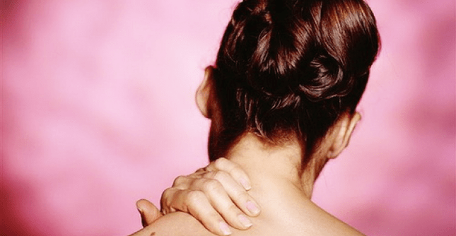
In the early stages of the disease, the pain can be explained by excessive fibrous ring and irritation of the posterior longitudinal ligament.The pain can be located in place in the back or neck, as well as in remote areas.With cervical osteochondrosis, the pain can be reflected in the back of the head, blade and interspace, shoulder and hand carriers.
Pain is associated with segmental muscle reflex spasm.This phenomenon has a protective nature and stabilizes the defined part of the spinal column.Over time, muscle contraction becomes an independent source of pain.When moved to the intervertebral hole, hernia squeezes the neighboring nerve roots.Radical pain has a character, permeable, clearly localized during nerve intrigue.Is associated with appropriate neurological manifestations:
- decreased sensitivity;
- reflex failure;
- Muscle weakness.
Disk degeneration violates the normal anatomical ratio between the components of the spinal column: discs, vertebrae, joints and ligaments.The gradual decrease in the height of the intervertebral disc leads to a change in articular connections and the formation of subluxation and vertebra displacements.This fact indicates the instability of the spinal column and reduces resistance to damage, which can lead to deterioration of osteochondrosis.
With age, the stability of the spine has been restored due to the formation of osteophytes, hypertrophy of articular processes, disk fibrosis, articular ligament thickening and capsules.The final stage of the pathological process is called spondylosis.Pain for this time comes down.
The main symptoms of cervical osteochondrosis
At the level of the cervical segments, the nerve roots and their arteries, the spinal cord and its vessels, and the spinal arteries may undergo compression.Spinal compression is possible due to posterior intervertebral hernia or posterior osteophytes.People with a narrow vertebral canal are particularly predisposed to this.With a hernia, signs of compression of cervical osteochondrosis develop quite quickly, and symptoms of the cerebrospinal fluvine current are softer.
It is very difficult to clinically discern the compression of the spinal cord with a tumor and hernia.The osteochondrosis of the cervical spine is manifested by a spastic paresis of the legs, disorders of sensitivity, pain and weakness in the hands.In some cases, signs of compression are combined with signs of ischemia of the spinal cord substance that arise as a result of compression of the spinal artery and radicular vessels.
Symptoms of damage to the anterior horns and ventral departments may suddenly develop with the inclusion of pyramid trails (blood supply to the anterial spinal arteries).Side spinal syndrome occurs: slow wing paresis, spastic leg paresis, damaged sphincter function.Sometimes symptoms of gross violation of deep sensitivity develop in the hands.After 2-3 weeks, the signs of a spinal cord begin to regress.In terms of pathological focus volume, we can say the severity of the remaining phenomena.
Cervical myelopathy
Myelopathy is a chronic istem for cervical osteochondrosis.A major role in the development of this syndrome is played by compression of blood vessels.The most characteristic is the loss of ventral parts of the side pillars and front horns.It is manifested by a spasticoatrophic paresis of the wings, a spastic paresis of the feet, a violation of the deep sensitivity of the feet (classic trio).
In a number of patients, the symptoms of lermita appear: a feeling of passing electrical discharge along the entire spine with the radiation of pain in the hands and feet when going.It is possible to develop amyotrophic lateral sclerosis in which there are no bulbous symptoms.
An important role in confirmation of myalopathy is played by MRI and CT, which detect the compression of the shell bag with osteophytes and a thick yellow bunch.
Signs of radical compression
Since the underlying discs are tired faster, spondillaarthrosis takes place in the respective segments.Osteophytes narrow the intervertebral holes and squeeze the roots (at the lumbar level most often a compression of disk hernia in the epidural space).When the head of the growth moves, the spine is damaged, which causes the formation of edema, which further narrows the intervertebral hole.Develop reactive inflammatory reactions.
Clinical Manifestations:
- C3 -Coreshok (under 2 cervical vertebrae, rarely occurs) - pain in the corresponding half of the neck, feeling of swelling of the tongue, a feeling of a throat coma;
- C4 -koreshok - pain in the proper flow of the shoulder, clavicle, trapezoidal muscle atrophy, a decrease in neck muscle tone (Roots 3 and 4 cervix increases the diaphragm tone, which leads to a relocation in the liver and the appearance of papyrous pain);
- C5 -Decor - neck pain and the outer surface of the shoulder, deltoid muscle hypotrophy;
- C6 -koreshok (one of the most common localizations) -The neck, blade, shoulder, radial surface of the forearm spreads in 1 finger, hands on the hands, the weakness of the two -headed muscle biceps;
- C7-Koreshok-pain spreads to 2-3 fingers, accompanied by paresthesia, weakness of the three-headed muscle;
- C8 -Coreshok -the pain lies on the surface of the forearm elbow on the 5th finger, accompanied by paresthesia.
Cervical reflex syndromes
Vertebral syndrome is manifested by acute cervical pain (bastard, cervix), less often chronic or subacute pain.The main sources of pain syndrome are a fibrous ring, posterior longitudinal ligament, joint capsule, tense muscles.Krivosheya is not as pronounced as the curvature of the spine at the lumbar level.
The pain is hurting, radiating to the back of the head.Intensify when driving or staying extended in a position.In palpation, the pain of the spinose processes and the capsules of the joints on the sore side (along the back surface of the neck of 3-4 cm is determined side than spicy processes).Inclusion in the process not only of the back but also the anterior muscles of the spine (anterial stairs, etc.) is characteristic.
Front staircase syndrome
Stairs muscle tension very often occurs with cervical osteochondrosis.The muscle is determined by the muscle in the form of a sternum in the form of stress, dense and increased in size compared to the healthy side.Due to tension, compression of supravichical vessels occurs, which is associated with pain and swelling in the hand, damaged sensitivity and motor activity (along the elbow nerve).The pain intensifies in a horizontal position.
Small chest muscle syndrome
The development mechanism is similar to the previous one.Compression of the frozen vascular radius occurs between the muscles and the shoulder bone (or process of the harvest) in conditions of adding the wrist.It is associated with chest pain, shoulder blade, hand.
Existing features are often regarded as heart pain with VSD (no acute attacks, the effect of taking nitroglycerin or sedatives is not, increased symptoms during movement and palpation of pain points).
The charming posterior syndrome
Disordishes distributing, vasomotor disorders that occur as a result of irritation of the sympathetic plexus of the vertebral artery are characteristic.Plexus branches are located in brain and skull tissue.It is clinically manifested by dizziness, a bell in the ear, spectacular disorders, anxiety.
Compression of vertebral arteries with osteophytes emanating from the spinal column joints, in combination with atherosclerotic damage to these vessels, is an important pathogenetic factor in the development of the insufficiency of the brain arteries and spinal cord.
cONcluSiON
In most cases, pain in the hands and neck is associated with cervical osteochondrosis.In some patients, the pain is caused by the hernia of the intervertebral disc, in others - osteophytes and arthrosis of the spine joints.Each of these options can lead to local or mirrored pain, radical syndrome and myelopathy.When examining patients with neck pain, it is necessary to exclude pathologies such as:
- spine tumors;
- epidural abscess;
- spondylitis;
- subarachnoid hemorrhage;
- meningitis;
- Hall abscess;
- stratification of carotid artery;
- Fracture of cervical vertebrae.